
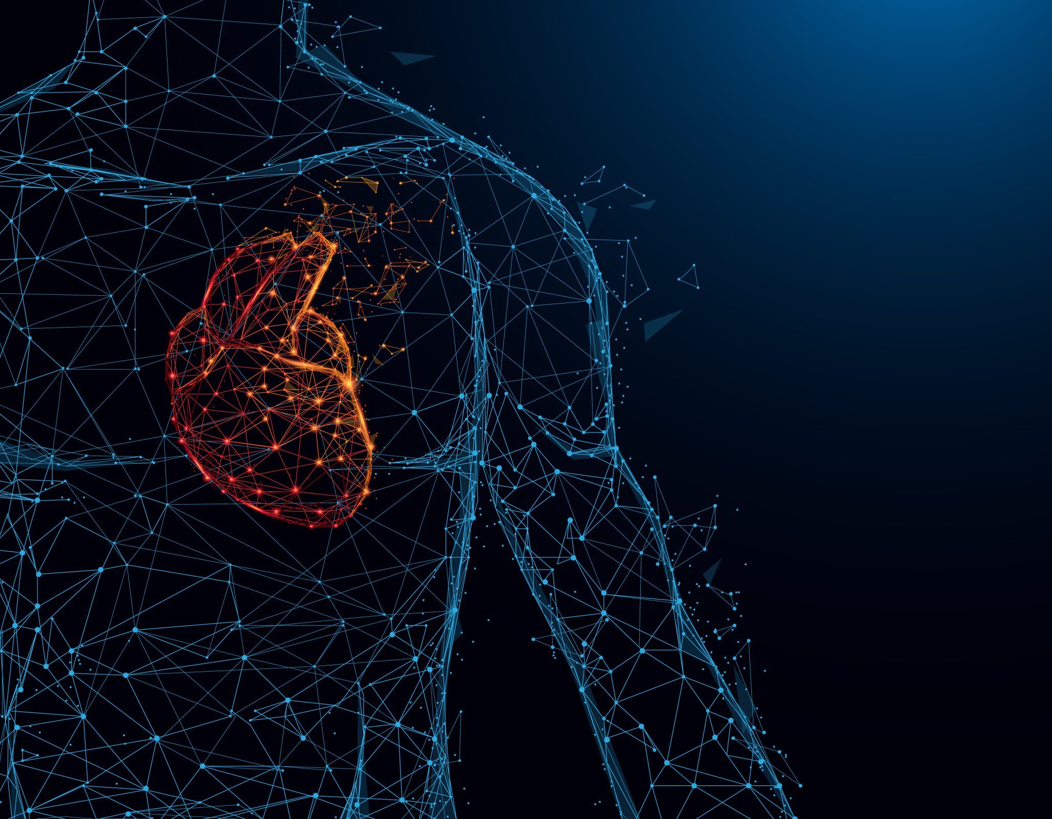
As fatty substances clog the arteries, the heart’s blood flow becomes obstructed, leading to coronary heart disease.
According to the World Health Organization, cardiovascular illnesses are the leading cause of mortality globally, taking more than 17.9 million lives yearly.
Around 14.4% of those with heart disease have coronary heart disease, which is a major source of worry for experts. When fatty substances clog the arteries leading to the heart, it results in coronary heart disease because the blood supply to the heart is cut off.
Studies link unhealthy lifestyles, poor diets, inactivity, cigarette use, and excessive alcohol intake to the obstruction of cardiac arteries.
Cardiomyocytes, the unique cells that make up the heart, become dysfunctional and necrotic as a result of diseases including coronary heart disease, which also destroy cardiac tissue.
Damaged cardiac tissue cannot regenerate back in humans. As an alternative, the organ develops scar tissue or the patient needs a heart transplant, which has its own set of difficulties.
Scientists from the Indian Institute of Technology, Guwahati, under the direction of Rajkumar P. Thummer, assistant professor in the department of biosciences and bioengineering, have now developed a method for reprogramming healthy skin cells from an adult into heart cells.

According to the experts, transforming cells from one form to another – a.k.a. cellular reprogramming – includes special proteins, called transcription factors, that modify the “expression of genes inside a cell and lead it to take on a new cellular identity”.
How does it function?
In a damaged heart, the irreversible loss of beating cells leads to the formation of scar tissue containing cardiac fibroblast [a type of cells]. In the field of cardiology, balancing this loss of cardiomyocytes is highly challenging, even in this modern medical world,” Dr Thummer told
“Therefore, our lab established a recombinant protein toolbox consisting of six potential cardiac transcription factors, which can convert these fibroblasts into functional cardiomyocytes.”
Six recombinant proteins make up the toolbox, according to Dr. Thummer: GATA4, MEF2C, TBX5, ETS2, MESP1, and HAND2. Each of these proteins is essential for the transformation of fibroblasts, a kind of cell that gives tissue structure, into cardiomyocytes.
While there has been research into creating heart cells from somatic cells (any cell of a live creature), this specific study looks for a more secure method.
“Fibroblasts are the most commonly use cell source for cellular reprogramming. The majority of the previous studies used an integrative approach based-cardiac reprogramming, which limits clinical applicability,” Dr Thummer said.
“Thus, we aimed to develop a safer approach for direct cardiac reprogramming, which can then be used to reprogram cardiac fibroblasts which form scar tissues after heart attack, into functional cardiomyocytes in the damaged heart.”
“Proteins are safer and necessary macromolecules in our diet. The advantage of recombinant proteins-based cell conversion is they work their miracle inside the nucleus. And eventually disappear over time without leaving behind their toxic waste, unlike their genetic counterparts. Thus, they are very safe for reprogramming compared to other approaches,” he explained.
The research team has stated an anticipation that the work would contribute to the development of functioning cardiomyocytes. That suited to a patient’s demands as well as improved cellular reprogramming efficiency.